PDF] Brain Tumor Segmentation of MRI Images Using Processed Image Driven U-Net Architecture
Por um escritor misterioso
Last updated 10 abril 2025
![PDF] Brain Tumor Segmentation of MRI Images Using Processed Image Driven U-Net Architecture](https://d3i71xaburhd42.cloudfront.net/c750894747d2b3f841de55922b2b68794295de27/7-Table3-1.png)
A fully automatic methodology to handle the task of segmentation of gliomas in pre-operative MRI scans is developed using a U-Net-based deep learning model that reached high-performance accuracy on the BraTS 2018 training, validation, as well as testing dataset. Brain tumor segmentation seeks to separate healthy tissue from tumorous regions. This is an essential step in diagnosis and treatment planning to maximize the likelihood of successful treatment. Magnetic resonance imaging (MRI) provides detailed information about brain tumor anatomy, making it an important tool for effective diagnosis which is requisite to replace the existing manual detection system where patients rely on the skills and expertise of a human. In order to solve this problem, a brain tumor segmentation & detection system is proposed where experiments are tested on the collected BraTS 2018 dataset. This dataset contains four different MRI modalities for each patient as T1, T2, T1Gd, and FLAIR, and as an outcome, a segmented image and ground truth of tumor segmentation, i.e., class label, is provided. A fully automatic methodology to handle the task of segmentation of gliomas in pre-operative MRI scans is developed using a U-Net-based deep learning model. The first step is to transform input image data, which is further processed through various techniques—subset division, narrow object region, category brain slicing, watershed algorithm, and feature scaling was done. All these steps are implied before entering data into the U-Net Deep learning model. The U-Net Deep learning model is used to perform pixel label segmentation on the segment tumor region. The algorithm reached high-performance accuracy on the BraTS 2018 training, validation, as well as testing dataset. The proposed model achieved a dice coefficient of 0.9815, 0.9844, 0.9804, and 0.9954 on the testing dataset for sets HGG-1, HGG-2, HGG-3, and LGG-1, respectively.
![PDF] Brain Tumor Segmentation of MRI Images Using Processed Image Driven U-Net Architecture](https://images.prismic.io/encord/57bd343a-7e54-4653-a716-f8fbd88d1afc_image+%284%29.png?auto=compress%2Cformat&fit=max)
Guide to Image Segmentation in Computer Vision: Best Practices
![PDF] Brain Tumor Segmentation of MRI Images Using Processed Image Driven U-Net Architecture](https://www.mdpi.com/computers/computers-10-00139/article_deploy/html/images/computers-10-00139-g003.png)
Computers, Free Full-Text
![PDF] Brain Tumor Segmentation of MRI Images Using Processed Image Driven U-Net Architecture](https://www.degruyter.com/document/doi/10.1515/jisys-2022-0206/asset/graphic/j_jisys-2022-0206_fig_001.jpg)
A novel deep learning-based brain tumor detection using the Bagging ensemble with K-nearest neighbor
![PDF] Brain Tumor Segmentation of MRI Images Using Processed Image Driven U-Net Architecture](https://www.researchgate.net/publication/349902492/figure/fig1/AS:1024432529215497@1621255156793/Proposed-tumor-segmentation-and-classification-architecture.png)
Proposed tumor segmentation and classification architecture
![PDF] Brain Tumor Segmentation of MRI Images Using Processed Image Driven U-Net Architecture](https://journals.sagepub.com/cms/10.1177/20552076221074122/asset/images/large/10.1177_20552076221074122-fig3.jpeg)
Magnetic resonance image-based brain tumour segmentation methods: A systematic review - Jayendra M Bhalodiya, Sarah N Lim Choi Keung, Theodoros N Arvanitis, 2022
![PDF] Brain Tumor Segmentation of MRI Images Using Processed Image Driven U-Net Architecture](https://media.springernature.com/m685/springer-static/image/art%3A10.1186%2Fs12859-022-04794-9/MediaObjects/12859_2022_4794_Fig2_HTML.png)
A state-of-the-art technique to perform cloud-based semantic segmentation using deep learning 3D U-Net architecture, BMC Bioinformatics
![PDF] Brain Tumor Segmentation of MRI Images Using Processed Image Driven U-Net Architecture](https://www.frontiersin.org/files/Articles/959667/fpubh-10-959667-HTML-r1/image_m/fpubh-10-959667-g001.jpg)
Frontiers Efficient framework for brain tumor detection using different deep learning techniques
![PDF] Brain Tumor Segmentation of MRI Images Using Processed Image Driven U-Net Architecture](https://html.scirp.org/file/1-9102840x4.png?20140121090614032)
Brain Tumor Segmentation of HGG and LGG MRI Images Using WFL-Based 3D U-Net
![PDF] Brain Tumor Segmentation of MRI Images Using Processed Image Driven U-Net Architecture](https://image.isu.pub/221203102411-2d295fd32a8526195049b137eb92dbb6/jpg/page_1_thumb_large.jpg)
Unet 3+ For Brain Tumor Segmentation : A Study by IRJET Journal - Issuu
![PDF] Brain Tumor Segmentation of MRI Images Using Processed Image Driven U-Net Architecture](https://www.medrxiv.org/content/medrxiv/early/2022/11/04/2022.11.03.22281923/F1.large.jpg)
Comparing 3D, 2.5D, and 2D Approaches to Brain Image Segmentation
![PDF] Brain Tumor Segmentation of MRI Images Using Processed Image Driven U-Net Architecture](https://journals.sagepub.com/cms/10.1177/20552076221074122/asset/images/large/10.1177_20552076221074122-fig1.jpeg)
Magnetic resonance image-based brain tumour segmentation methods: A systematic review - Jayendra M Bhalodiya, Sarah N Lim Choi Keung, Theodoros N Arvanitis, 2022
![PDF] Brain Tumor Segmentation of MRI Images Using Processed Image Driven U-Net Architecture](https://media.springernature.com/m685/springer-static/image/art%3A10.1186%2Fs12859-022-04794-9/MediaObjects/12859_2022_4794_Fig1_HTML.png)
A state-of-the-art technique to perform cloud-based semantic segmentation using deep learning 3D U-Net architecture, BMC Bioinformatics
![PDF] Brain Tumor Segmentation of MRI Images Using Processed Image Driven U-Net Architecture](https://d3i71xaburhd42.cloudfront.net/d12c02378276e1fb4f8dd00fd8b25e9761866a56/3-Figure1-1.png)
PDF] Attention Gate ResU-Net for Automatic MRI Brain Tumor Segmentation
![PDF] Brain Tumor Segmentation of MRI Images Using Processed Image Driven U-Net Architecture](https://d3i71xaburhd42.cloudfront.net/420f3f1078a6d8e0696572c032877079286051c6/3-Figure1-1.png)
PDF] Brain Tumor Segmentation Using Convolutional Neural Networks in MRI Images
![PDF] Brain Tumor Segmentation of MRI Images Using Processed Image Driven U-Net Architecture](https://media.springernature.com/m685/springer-static/image/art%3A10.1186%2Fs12859-021-04347-6/MediaObjects/12859_2021_4347_Fig1_HTML.png)
MRI-based brain tumor segmentation using FPGA-accelerated neural network, BMC Bioinformatics
Recomendado para você
-
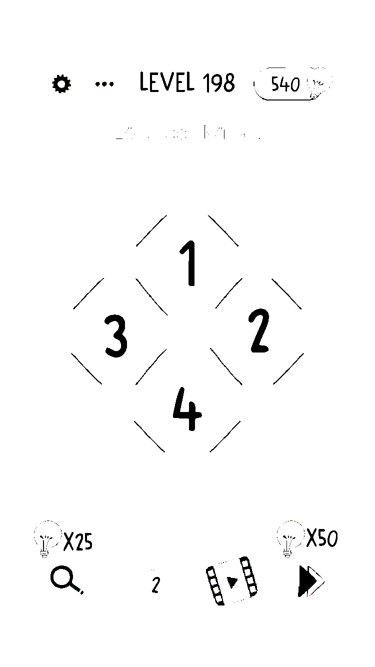 Brain Test: Level 191 bis Level 200 - Lösungen10 abril 2025
 -
Brain test level 191 - Cuaca hujan membuatku muram #jawaban #Games #br10 abril 2025
-
 Brain Test 4 Level 191 My cat must reach to her food Answers and Solutions10 abril 2025
 -
 brain test nível 19110 abril 2025
 -
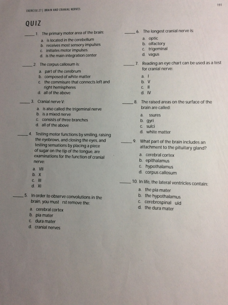 Solved 191 EXERCISE 27, BRAIN AND CRANIAL NERVES QUIZ 1.10 abril 2025
 -
 Some speech disorders. Journal of Neurology, Neurosurgery & Psychiatry10 abril 2025
 -
 BRINCADEIRA DE GATOS 😂😂😂😂😂😂😂😂 #HUMORPET em 202310 abril 2025
 -
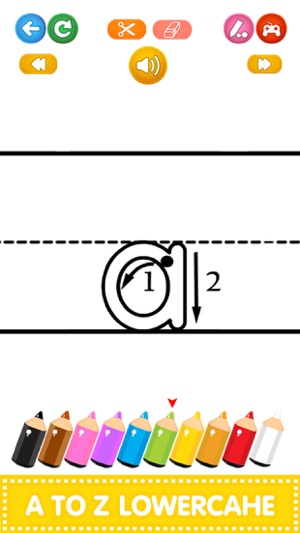 ABC 123 Learn to Write Letters on the App Store10 abril 2025
 -
Pet doll for Android - Download the APK from Uptodown10 abril 2025
-
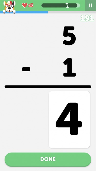 Math Learner: Learning Game, Apps10 abril 2025

você pode gostar
-
 NEMO10 abril 2025
 -
![Warder] Pure RETALIATION, REFLECT, INVINCIBLE, 100k+ DAMAGE, Hardcore Ultimate 100% Done (SF Gear) - Classes, Skills and Builds - Crate Entertainment Forum](http://i.imgur.com/uYuUEuI.png) Warder] Pure RETALIATION, REFLECT, INVINCIBLE, 100k+ DAMAGE, Hardcore Ultimate 100% Done (SF Gear) - Classes, Skills and Builds - Crate Entertainment Forum10 abril 2025
Warder] Pure RETALIATION, REFLECT, INVINCIBLE, 100k+ DAMAGE, Hardcore Ultimate 100% Done (SF Gear) - Classes, Skills and Builds - Crate Entertainment Forum10 abril 2025 -
 Jogo De Tabuleiro Infantil E Adulto Xadrez & Dama Pais & Filhos no Shoptime10 abril 2025
 -
 The Last of Us 2 - Joel pushes Ellie into water10 abril 2025
 -
 Boots launches a premium advent calendar with £450 worth of beauty10 abril 2025
 -
 One Piece WCF World Collectable Figure beasts Pirates 3 set King Queen Jack New10 abril 2025
 -
 Quebra-cabeça Cognitivo Hot Wheels10 abril 2025
 -
 Tuck Everlasting: 6 Vocabulary Crosswords by Sections—Unique!10 abril 2025
 -
 Door with Black Angry Scary Monster Face. Stock Photo - Image of10 abril 2025
 -
 Jogar paciência do Windows – Comentários de um Mundo Apple10 abril 2025