Morphology of Leydig cells in the testes after in vivo MCP-1 treatment.
Por um escritor misterioso
Last updated 12 abril 2025


Stem Leydig cells: Current research and future prospects of regenerative medicine of male reproductive health - ScienceDirect
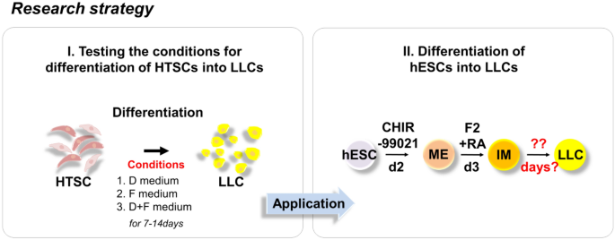
Rapid Differentiation of Human Embryonic Stem Cells into Testosterone-Producing Leydig Cell-Like Cells In vitro

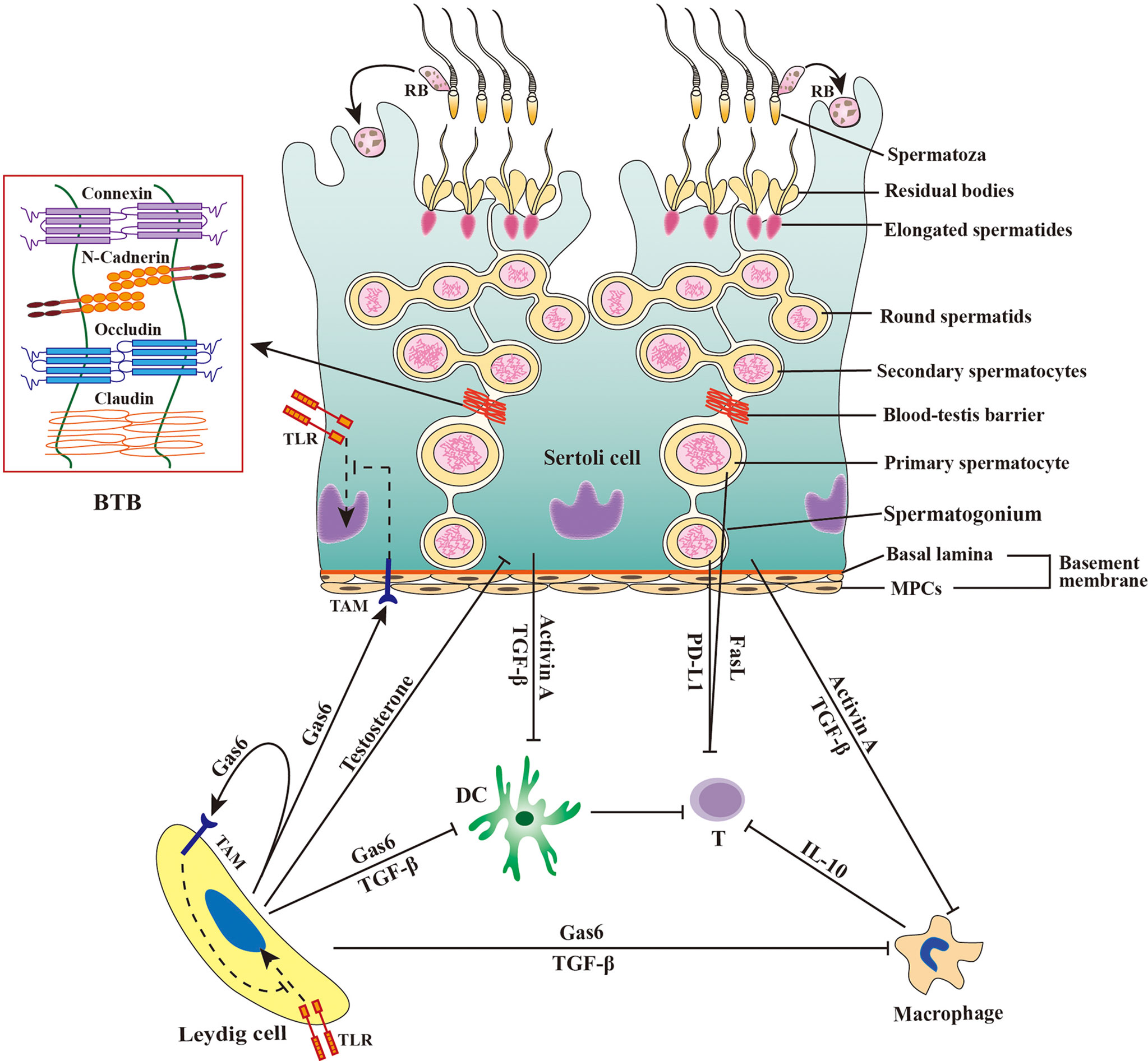
Prenatal exposure to bisphenol AF induced male offspring reproductive dysfunction by triggering testicular innate and adaptive immune responses - ScienceDirect
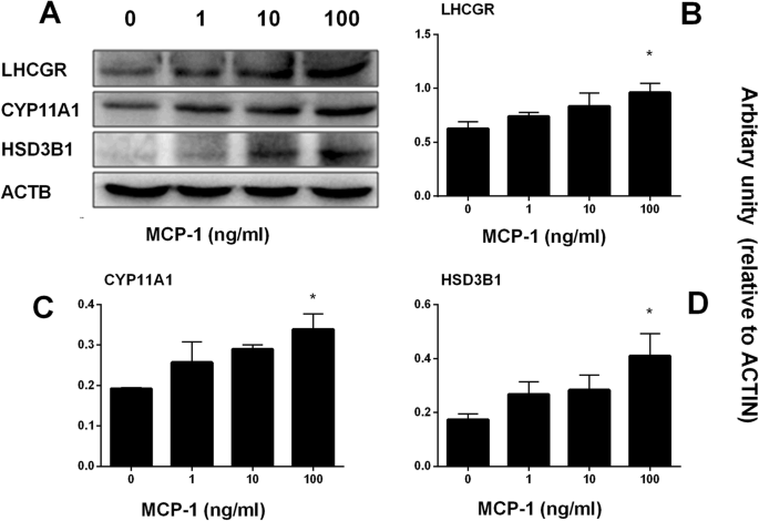
Monocyte Chemoattractant Protein-1 stimulates the differentiation of rat stem and progenitor Leydig cells during regeneration, BMC Developmental Biology
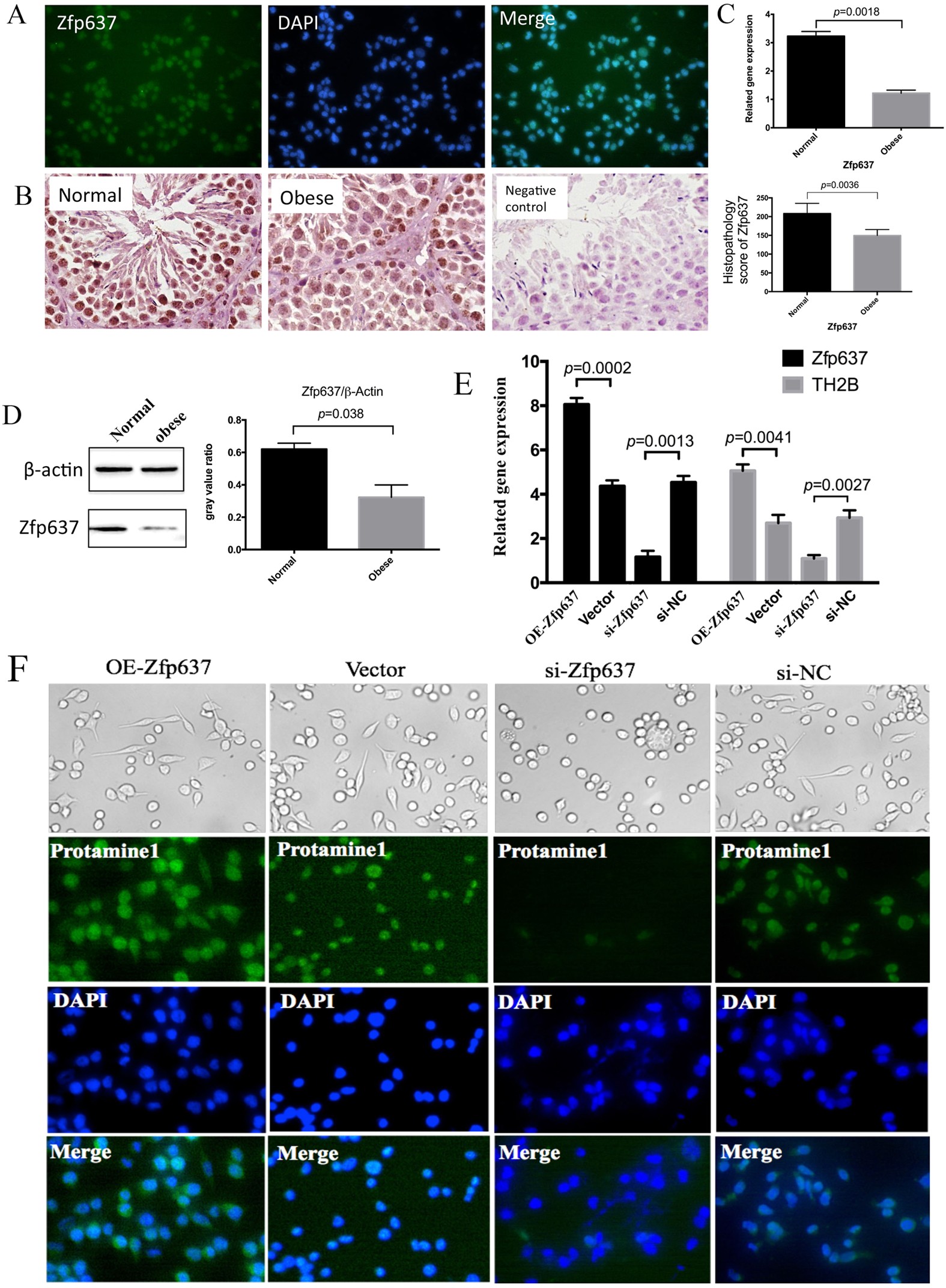
IL-6 mediates differentiation disorder during spermatogenesis in obesity-associated inflammation by affecting the expression of Zfp637 through the SOCS3/STAT3 pathway
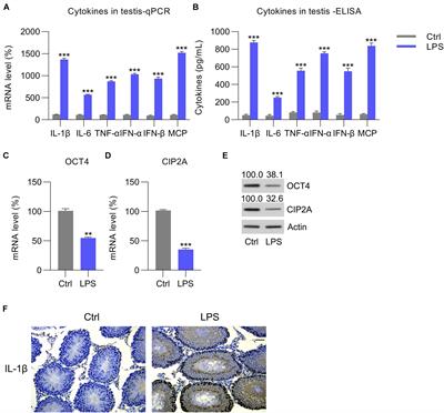
Frontiers OCT4 Represses Inflammation and Cell Injury During Orchitis by Regulating CIP2A Expression
Frontiers Viral tropism for the testis and sexual transmission

Therapeutic application of Sertoli cells for treatment of various diseases - ScienceDirect

Cell Type-Specific Expression of Testis Elevated Genes Based on Transcriptomics and Antibody-Based Proteomics

Ibuprofen and Leydig cell steroidogenic function. (A–C) Representative

The Sertoli cell: one hundred fifty years of beauty and plasticity - França - 2016 - Andrology - Wiley Online Library

Testicular torsion in vivo models: Mechanisms and treatments - Minas - 2023 - Andrology - Wiley Online Library

Testicular macrophages are recruited during a narrow time window by fetal Sertoli cells to promote organ-specific developmental functions
Recomendado para você
-
 Teste de Velocidade da Internet Vivo12 abril 2025
 -
 Teste Palográfico: da técnica à prática - Grupo Educativa12 abril 2025
 -
 Sobre Um Fundo Vermelho Vivo Um Cartão De Cor De Madeira Leve Com Um Teste De Massa Ilustração Stock - Ilustração de palavra, mensagem: 23630853212 abril 2025
 -
 ROWCES Caneta multímetro digital NCV AC/DC Voltímetro Resistência do ohmímetro Capacitância Frequanecy Teste de linha ao vivo Testador de luz de fundo de LCD portátil de 4000 contagens com retenção12 abril 2025
 -
 Tablet vivo Pad é encontrado em testes no Geekbench12 abril 2025
 -
![Vivo Fibra 100MB - Teste de velocidade Vivo de 100MB [2018]](https://i.ytimg.com/vi/L-YmHTsWhFc/hqdefault.jpg) Vivo Fibra 100MB - Teste de velocidade Vivo de 100MB [2018]12 abril 2025
Vivo Fibra 100MB - Teste de velocidade Vivo de 100MB [2018]12 abril 2025 -
Teste de Transmissão ao vivo12 abril 2025
-
72º Curso: Teste Palográfico na Avaliação da Personalidade - Transmissão ao Vivo12 abril 2025
-
 Estamos Chegando a Imagem Escrita De Teste Ao Vivo Com Design Floral Ilustração Stock - Ilustração de arte, escrito: 21186803612 abril 2025
 -
 Beleza blogger teste escova de sombra para os olhos na frente da câmera, gravação de vídeo tutorial de maquiagem, streaming ao vivo em casa.12 abril 2025

você pode gostar
-
 Tattoo uploaded by Filipe Lopes • Xadrez da vida! #xadrez #chess12 abril 2025
 -
 True Love Waits (Short 2017) - IMDb12 abril 2025
 -
 I am trying to make a death effect - Scripting Support - Developer Forum12 abril 2025
 -
 Moto motorizada infantil +23 anúncios na OLX Brasil12 abril 2025
 -
 Folding Magnetic International Chess Magnetic International Chess12 abril 2025
 -
 Stream Stranger Things Season 4 Episode 9 Song Running Up That Hill (EP9 Remix Version) by bruh deimos💀12 abril 2025
 -
 menhera chan kawaii surprise | Sticker12 abril 2025
 -
 Easily 3 Star the Goblin King Challenge (Clash of Clans)12 abril 2025
 -
 Resenha, FUUTO PI, Episódios 1 e 212 abril 2025
 -
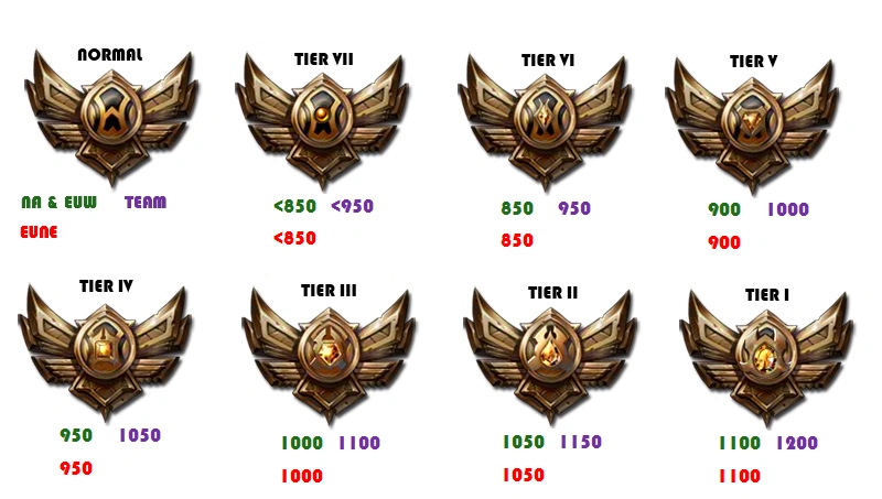 Elo rating system, League of Legends Wiki12 abril 2025