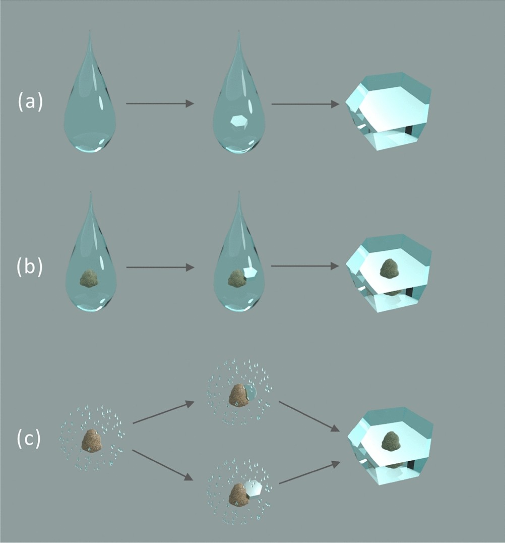
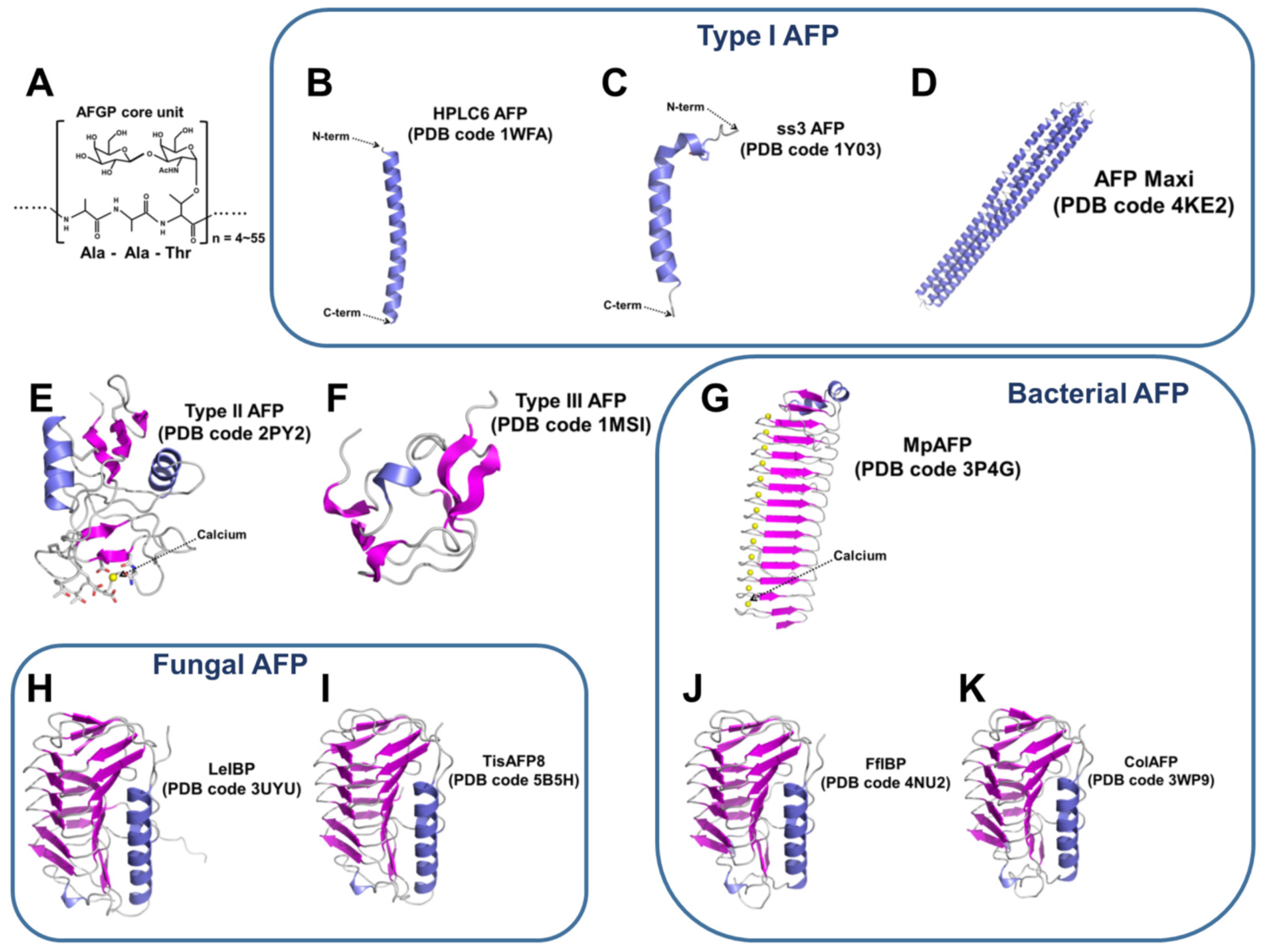
Electron microscopy and calorimetry of proteins in supercooled
Por um escritor misterioso
Last updated 26 abril 2025
Polymers, Free Full-Text

Chilled proteins and 3-D images: The cryo-electron microscopy
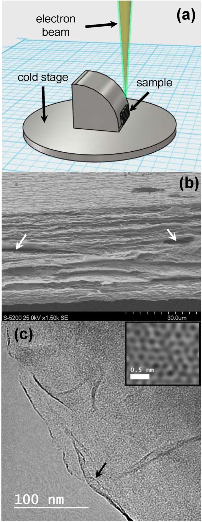
In-Situ ESEM and EELS Observation of Water Uptake and Ice

Glass transition and dynamics in BSA–water mixtures over wide

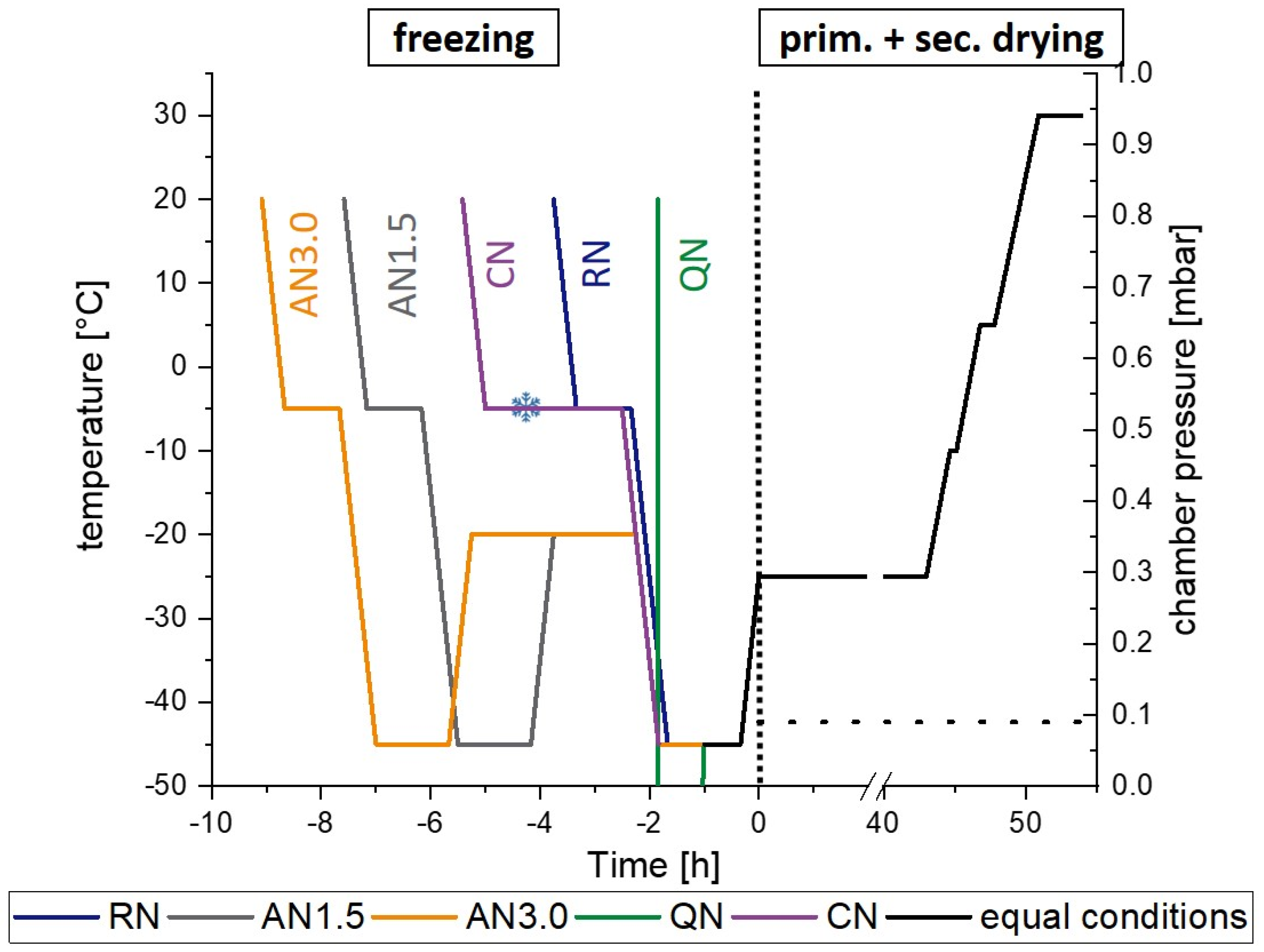
Freeze-dried cake structural and physical heterogeneity in
Pharmaceutics, Free Full-Text
Marine Drugs, Free Full-Text
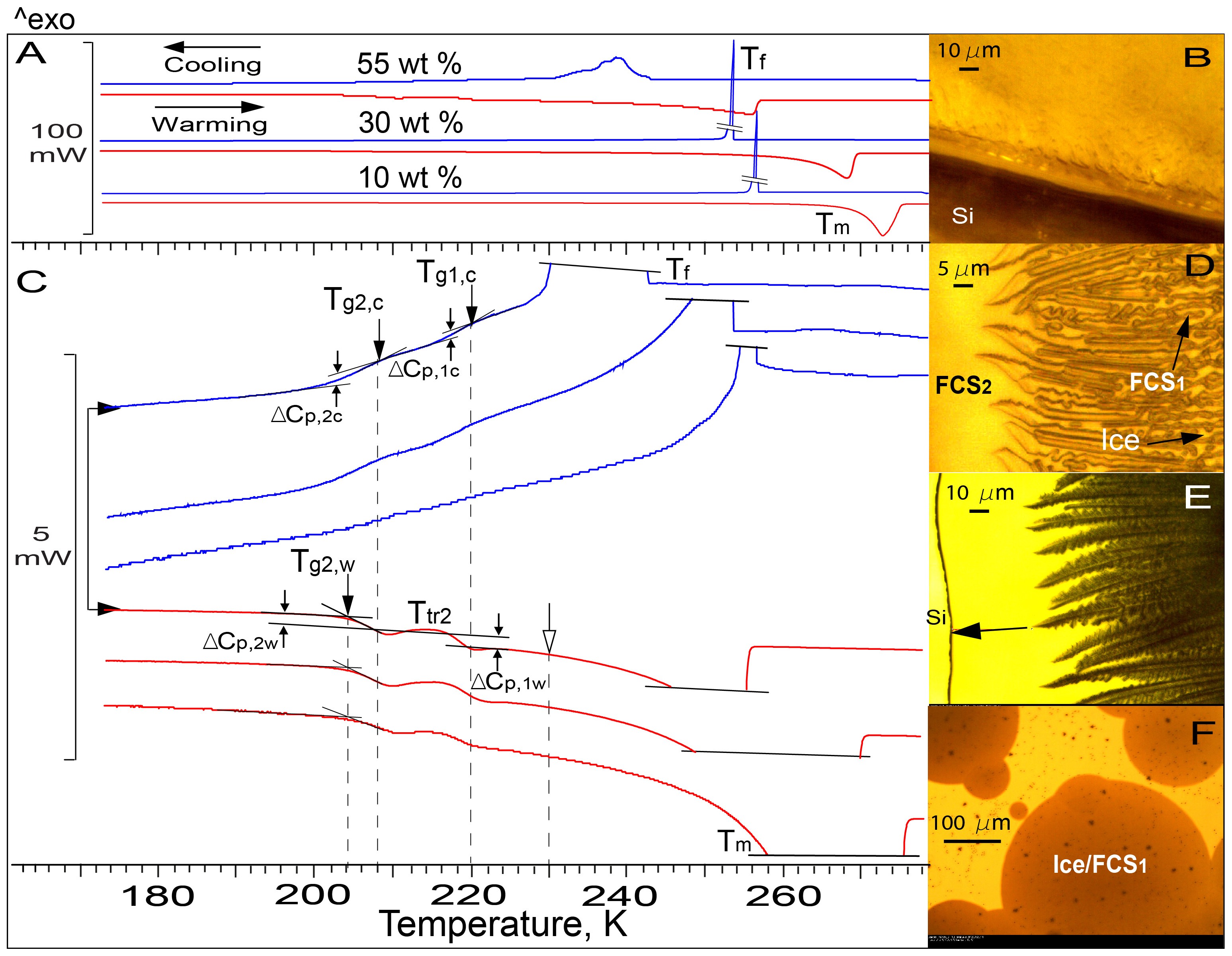
Visualization of Freezing Process in situ upon Cooling and Warming

Full article: Protein Stability During Freezing: Separation of

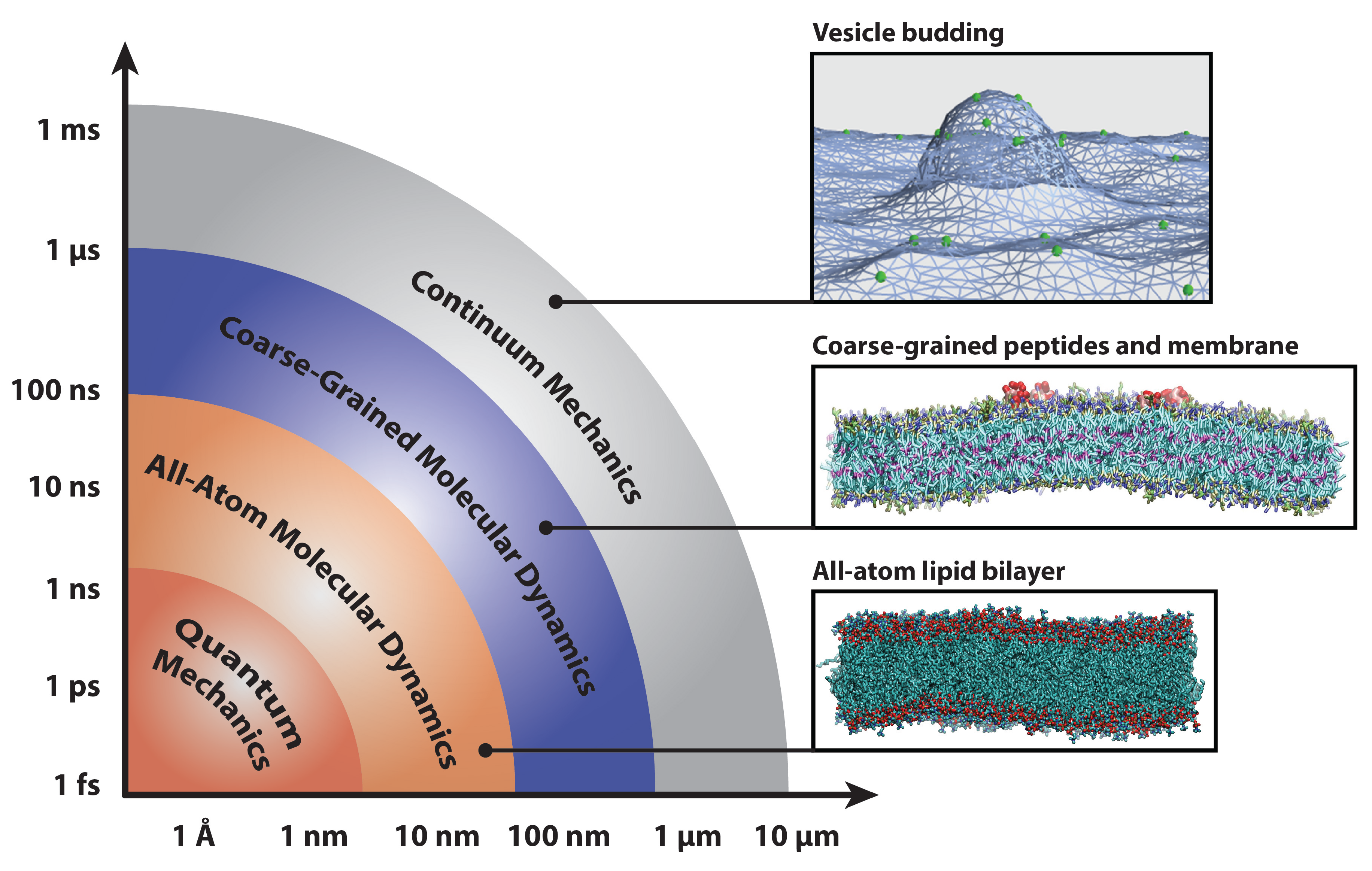
Confined Water as Model of Supercooled Water

Ice nucleation spectra of Pseudomonas strains submitted to
Electron microscopy and calorimetry of proteins in supercooled

Full article: Protein Stability During Freezing: Separation of
pH dependence of frozen fractions at six different pH values for

Types of differential scanning calorimeters: (a) heat-flux
Recomendado para você
-
 World Cube Association26 abril 2025
 -
 2023 CONCERT FOR CAUSE – DECEMBER 14TH – KRVB26 abril 2025
 -
 About Writers' Club Amino26 abril 2025
 -
 Resume.pdf26 abril 2025
 -
 Synthetic polyurethane nanofibrous membrane with sustained26 abril 2025
 -
 Alliance defeat Team Empire, clinch WCA 2015 LAN spot Dota Blast26 abril 2025
 -
 Wildcard Gaming - Leaguepedia League of Legends Esports Wiki26 abril 2025
 -
 Watercolor Artist Fall 2022 Digital Edition26 abril 2025
 -
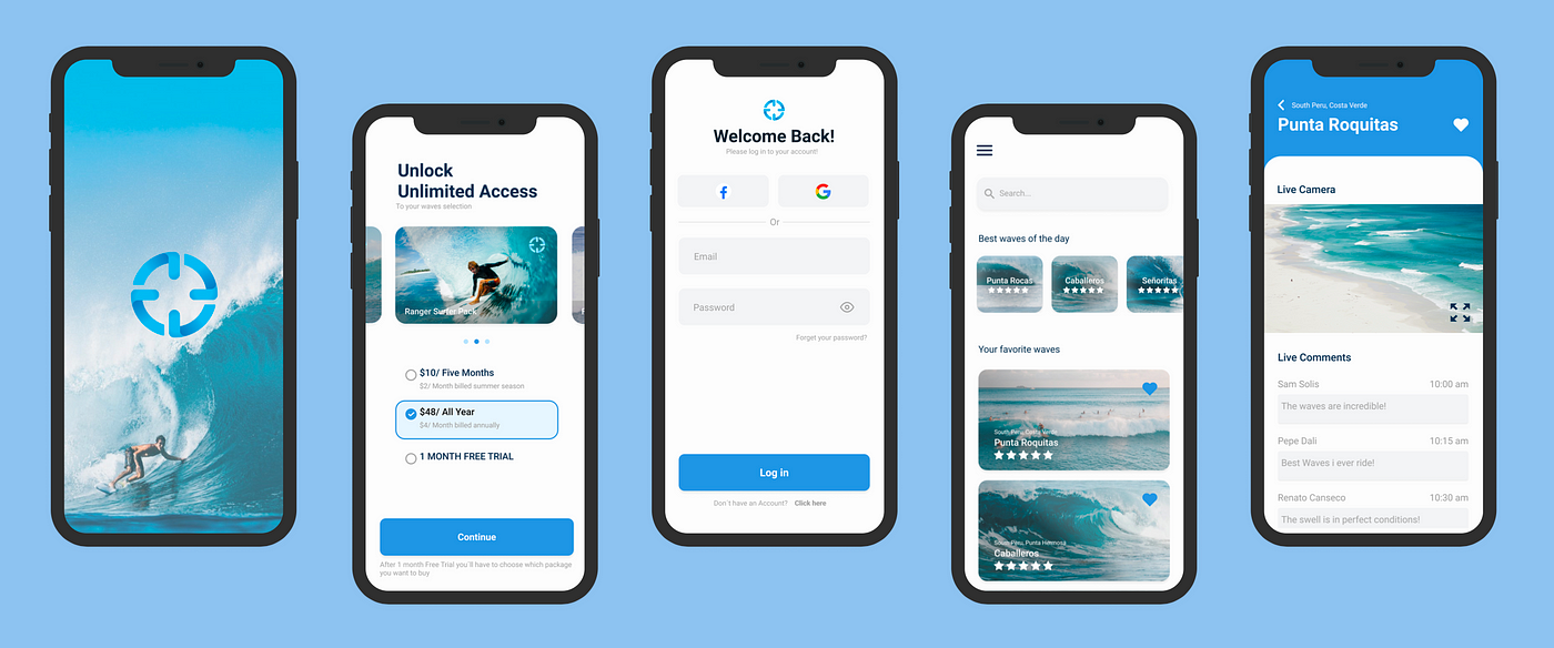 UI/UX Case Study: Redesign Mis Olas (My Waves) App, by Valeriadavila26 abril 2025
 -
Digital Rage Podcast on Spotify26 abril 2025

você pode gostar
-
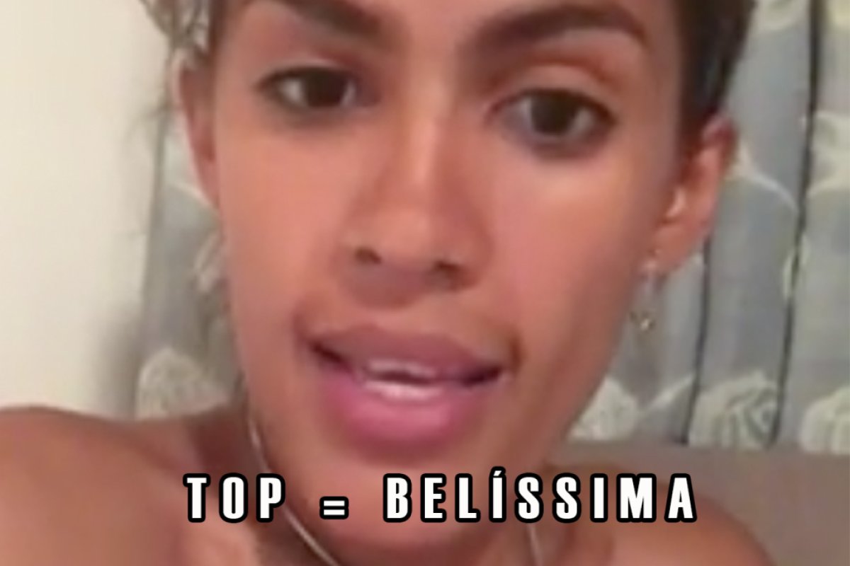 Esta brasileira ensina o significado da palavra “top” e é26 abril 2025
 -
 Enjoying the Journey a podcast by Scott Pauley / Enjoying the26 abril 2025
 -
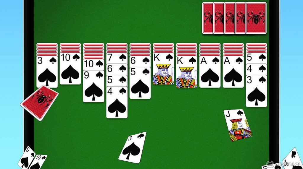 Spider Solitaire PC Version - Free Card Game Download26 abril 2025
 -
 Anime Bleach HD Wallpaper by Kazuaki26 abril 2025
 -
 Google Play Pass: conheça o novo serviço de assinatura de games26 abril 2025
 -
 Metaverso: como funciona, tecnologias, como entrar e investir - FIA26 abril 2025
 -
Asrai Unit Pack (Wood Elves) - Skymods26 abril 2025
-
 Observation Inf Range Blox Fruits Script Download 100% Free26 abril 2025
 -
 Perguntas sobre msica quiz26 abril 2025
 -
![Linhas De Xadrez Textura Colorida Vetor EPS [download] - Designi](https://www.designi.com.br/images/preview/11341240.jpg) Linhas De Xadrez Textura Colorida Vetor EPS [download] - Designi26 abril 2025
Linhas De Xadrez Textura Colorida Vetor EPS [download] - Designi26 abril 2025